loose body histology
Results The 84 loose bodies included 48 chondral loose bodies type I 26 osteochondral loose bodies type II and 10 osseous loose bodies type III. BOX 83-1 Classification of Loose Bodies.

Trachea Histology Lamina Propria Hyaline Cartilage Respiratory Epithelium Respiratory Respiratory System Pathology Study
A loose body in a joint can be attributed to various factors such as osteochondritis dissecans osteochondral fracture synovial chondromatosis or fracture of osteophytes in osteoarthritis.

. The 26 osteochondral loose bodies type II could be subdivided into 8 composed of cartilage with enchondral ossification type IIa 11 consisting of mature bone covered by cartilage without. Osteochondral loose body consisting mainly of central cartilaginous material with focal endochondral ossification and covered with a layer of fibrotic synovial membrane. The conservative care was discontinued at this point and the loose bodies were removed.
No clumped atypical chondrocytes. May form if portion of articular cartilage detached cartilage or cartilagebone within joint space with necrotic calcified centers may become attached to synovial membrane revascularize and convert to viable bone breaks off. Loose bodies are usually asymptomatic 1.
A histopathological analysis of 119 surgically excised loose bodies revealed that the cases could be separated into three categories. It is well known that loose bodies grow from proliferation of cartilage without blood supply in the joint cavity and that enchondral ossification is able to develop only under the condition of having a blood supply. Mucocele traumatized hemangioma pyogenic granuloma.
Has the tide mark of articular cartilage has evidence of prior structure. Peritoneal loose bodies are formed by the torsion and autoamputation of epiploic appendages. We histologically examined 84 loose bodies and 9 related lesions synovial membrane nodules surgically removed from 24 joints of 24 patients with osteoarthrosis.
Acute torsion produces epiploic appendagitis which is normally self limiting. No unevenly distributed chondrocytes. When the loose body is just cartilage or in the case of synovial chondromatosis an MRI is the best non-radiation diagnostic.
Up to 10 cash back Histologically based analyses of the nature and origin of loose bodies occurring in osteoarthrosis have been few and further study is warranted. With special reference to their pathology and etiology A. Synovial Chondromatosis is a proliferative disease of the synovium associated with cartilage metaplasia that results in multiple intra-articular loose bodies.
Blood vessel rich - key element proliferation of fibroblasts - key element inflammation - especially lymphocytes plasma cells common - evidence of erosionulceration. 3 loose bodies due to joint surface disintegration. Histologic classification of loose bodies in osteoarthrosis.
As synovial membrane nodules were also classified to the same types as loose bodies. First described in the knee by Ambroise Paré 32 in 1558 loose bodies occur in the elbow with a frequency second only to that in the knee. May form if portion of articular cartilage breaks off.
In most cases the traumatic loose body has a bone chip with it or a big chunk of cartilage easily seen on an x-ray. The best way to see the location and character of a loose body is with an MRI. Histopathological examination of the loose bodies showed multiple subsynovial cartilaginous nodules TableFig 2 and mild to moderate cellular atypia in the shape and size of the chondrocytes of the subsynovial cartilaginous nodules TableFig 3.
Of note the popliteus bursa is a normal fluid-filled and synovial-lined structure along the extra-articular tendon and musculotendinous junction which on occasion contains. Epiploic appendages are suscepitible to torsion due to their narrow pedicle. The classification of loose bodies in human joints.
A loose body is typically diagnosed with an x-ray. 1 loose bodies due to synovial osteochondromatosis. Normally loose body is nourished by synovium and continues to grow has a tree ring appearance.
The condition usually presents in patients between 30 and 50 years of age with localized joint pain stiffness and swelling. Diagnosis is made on radiographs in late disease but MRI studies may be required in. 33 As in other joints it is sometimes difficult to distinguish with certainty between ossification centers and acquired lesions Box 83-1.
2 loose bodies due to osteochondral fracture. Attenuated synovial cells are present on the surface Haematoxylin and Eosin stained section original magnification 5 objective. A study of loose bodies composed of cartilage or of cartilage and bone occuring in joints.
Has the tide mark of articular cartilage has evidence of prior structure. Assessment of the popliteal hiatus on sagittal images is important as pathology such as meniscal flaps and loose bodies occasionally extend into this area Figure 5. Normally loose body is nourished by synovium and continues to.

Nervous Tunic Histology Slide Pigmented Epithelium Google Search Medical Laboratory Science Medical School Studying Biology Labs
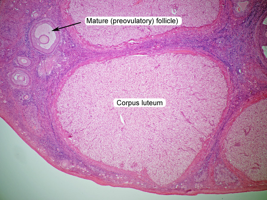
Corpus Luteum Medical Studies Corpus

Dense Connective Tissue Microanatomy Web Atlas Gwen V Childs Ph D Tissue Types Loose Connective Tissue Microscopic Cells

Histological Skin Structure Diagram Skin Structure Loose Connective Tissue Subcutaneous Tissue

Siu Som Histology Intro Human Anatomy And Physiology Stratified Squamous Epithelium Anatomy And Physiology

Loose Areolar Connective Tissue Histology Human Anatomy And Physiology Histology Slides Anatomy And Physiology

Anatomia Biologia Fisiologia

Lamina Propria Histology Loose Connective Tissue Medical Assistant Student Human Tissue

Thin Skin Integumentary System Medical School Studying Human Anatomy And Physiology

Pin On Tongue

Pin On Histologia

Pin On Chapter 5 Histology

Kinds Of Epithelial Tissues Stratified Squamous Epithelium Anatomy And Physiology Tissue Biology

Pin On Nursing School Study Tips And Guides

Histology Integumentary System At University Of Tasmania Studyblue Integumentary System Medical School Studying Anatomy And Physiology

Siu Som Histology Gi Histology Slides Medical Laboratory Science Medicine Notes

Stomach Histology Stomach Histology Slide Loose Connective Tissue Histology Slides Human Anatomy And Physiology

The Respiratory System Veterinary Anatomy Histology Respiratory System Loose Connective Tissue Respiratory

Pin On Pathology


Comments
Post a Comment